

A recent Nature study identifies Sst-Chodl neurons that actively promote sleep onset in mice, validating the complex biological shift from alertness to rest.





On September 9, 2026, researchers published a study in Nature reporting that a rare population of cortical neurons can promote sleep and synchronize activity across broad areas of the neocortex in mice. The research was led by Renata Batista-Brito, PhD, whose laboratory conducted the work while based at Albert Einstein College of Medicine before she later joined the Icahn School of Medicine at Mount Sinai.
The study focused on an exceptionally small population of cells known as Sst-Chodl neurons. The researchers described these cells as making up roughly 0.2 percent of cortical neurons. This extreme rarity makes their influence on overall brain state particularly notable for sleep researchers. They represent a highly specialized biological circuit operating within a much larger neural network.
Unlike many inhibitory neurons that primarily communicate with nearby cells, these specialized neurons send signals over long distances. This wide reach potentially allows them to coordinate activity across large cortical regions. That structural ability to cast a wide signal net appears crucial for their specific function. It suggests a mechanism designed for broad synchronization rather than isolated local control.
In alert mice, these specific neurons remained largely quiet during periods of wakefulness. The researchers observed that their cellular activity steadily increased as the animals became drowsy and began to enter deep non-rapid eye movement sleep. This targeted activation closely coincided with the emergence of slow and synchronized cortical rhythms. These slow rhythms are traditionally associated with deep and restorative rest.
To test their capability directly, the researchers experimentally activated the Sst-Chodl neurons. This experimental activation made electrical activity across the neocortex slower and significantly more synchronized. It produced electrical patterns closely resembling normal sleep-related cortical activity. Furthermore, this targeted activation caused the mice to fall asleep more readily and increased the total amount of time they spent sleeping.
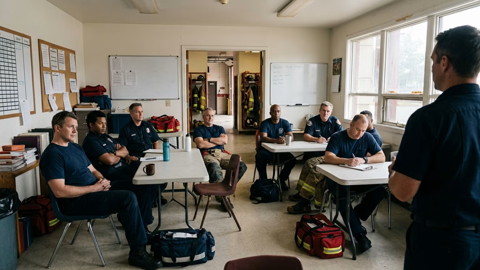
The study offers a highly useful biological framework for thinking about sleep onset as an active process. This transition from wakefulness into sleep represents a distinct and critical phase of physical rest. For emergency-service professionals, understanding this physiological distinction carries immediate practical value. Personnel frequently finish a late or overnight duty period while still physiologically alert.
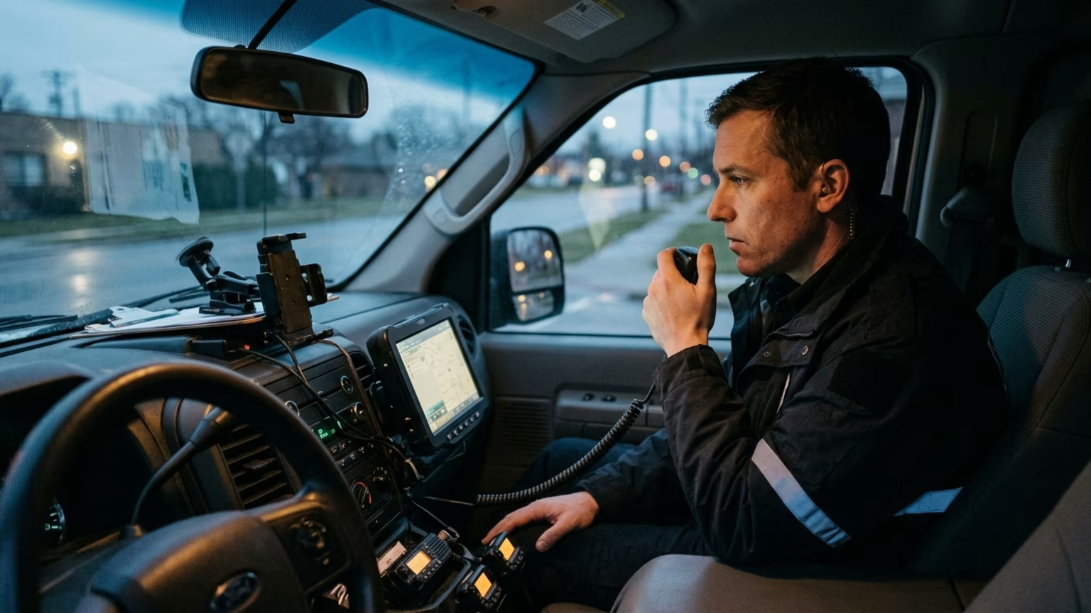
First responders regularly deal with irregular light exposure and high-stress activity right up to the end of a shift. A person may have a sufficient block of time scheduled for recovery, but they still struggle with the transition from duty-related alertness to sleep. This new research points out that sleep onset is a specific biological transition rather than a simple passive shutdown. By focusing on how the brain actively transitions out of alertness, the study validates the severe friction many professionals experience.
While the study did not test irregular duty schedules, it highlights why a protected transition period matters. Acknowledging that the brain requires active coordination to initiate sleep helps explain why moving directly from shift duties to bed often fails. Recognizing this active biology is crucial for anyone attempting to build a reliable sleep plan for nights and rotating shifts. Personnel cannot simply force their brains to power off without addressing this active physiological transition.
This perspective reinforces the need for structured recovery protocols after long and demanding tours. When sleep onset is viewed as an active requirement for cortical synchronization, the difficulty of winding down after work makes biological sense. It separates the frustration of lying awake from personal failure, framing it instead as an incomplete biological transition that requires deliberate management.
These findings fundamentally challenge the traditional view that the cortex mainly follows sleep signals generated elsewhere in the brain. For decades, the prevailing scientific model suggested that deeper brain structures dictated sleep states and subsequently imposed them on a passive cortex. The Nature study, titled "Neocortical long-range inhibition promotes cortical synchrony and sleep," introduces a significantly more dynamic picture of mammalian sleep regulation.
Batista-Brito noted that the traditional view of the cortex is as a passive follower during sleep. Instead, the recent study suggests that specialized cortical circuits can actively help initiate and organize the complex transition into sleep. She pointed out that these rare neurons did not simply become active after sleep had already begun. She explicitly stated that they can actually help drive the transition toward sleep.
This research does not imply that the cortex works entirely alone or replaces deeper sleep-regulating systems. The more measured interpretation is that natural sleep likely emerges from interaction between specialized deep-brain systems and active cortical circuits. Researchers proposed that these specific neurons might help translate accumulated sleep pressure into sleep onset. However, Batista-Brito clearly described that possibility as an untested hypothesis rather than an established conclusion.
This research represents foundational neuroscience and does not provide an immediate, ready-to-use intervention for shift workers. There is currently no evidence that emergency professionals can improve sleep by directly targeting Sst-Chodl neurons. Neither the study nor the available coverage reports experiments involving firefighters or police officers. Data is also missing for paramedics, night-shift workers and personnel on rotating schedules.
However, the findings do highlight the biological complexity of transitioning from wakefulness to rest. First responders should deliberately separate their approach to sleep onset from their approach to total sleep opportunity. A structured transition routine helps properly manage the shift from duty-related alertness to baseline physical recovery. Since the biological mechanism of falling asleep requires active neural coordination, personnel should focus on environments and daily habits that support this transition.
For instance, maintaining structured fueling and caffeine strategies can prevent unnecessary chemical interference with the brain's natural wind-down signals. Professionals must also keep established clinical sleep-care pathways firmly in place. The study does not establish a new medical or behavioral intervention based on Sst-Chodl neurons. Persistent difficulty falling asleep after shifts should consistently prompt a thorough discussion with a qualified healthcare professional.
This remains particularly critical when personnel experience excessive daytime sleepiness or witness breathing interruptions. Tracking overall sleep regularity on rotating shifts continues to be a recommended baseline practice for all frontline personnel. Acknowledging that the cortex plays an active role in sleep initiation should encourage professionals to optimize their sleep environments carefully. Managing room temperature, securing absolute darkness and reducing ambient noise all help support the brain as it attempts to orchestrate these complex sleep transitions.
The researchers observed that these specific neurons appear to have been evolutionarily conserved across hundreds of millions of years. Related cells have been successfully identified across amphibians, reptiles and humans. While that extensive evolutionary conservation may indicate an important biological role, it does not automatically establish clinical relevance. It does not prove that human Sst-Chodl neurons perform the exact same sleep-promoting function demonstrated in the mouse model.
Future research will carefully need to determine if the same circuit operates similarly in human populations. The available coverage does not report the number of mice or detailed activation protocols. It also omits quantitative sleep-onset latency, effect sizes and statistical significance values. Full control-condition details are similarly missing from the available reports.
These omissions make it inappropriate to claim a specific sleep benefit at this early stage. Scientists also need to investigate whether this specific circuit responds directly to prolonged wakefulness and irregular daily schedules. It remains completely unknown if occupational sleep disruption fundamentally changes Sst-Chodl activity. Researchers do not yet know if the neural circuit becomes physically impaired after extended duty hours or chronic sleep debt.
The most important next questions involve whether changes in this circuit contribute to sleep problems linked to specific systemic diseases. The recent finding provides a specific biological circuit for future investigation rather than a definitive current treatment plan. Until more human translation and clinical effectiveness data fully emerges, professionals evaluating options for first responder sleep and recovery should continue relying on established science.
Understanding the biological mechanisms of sleep onset clarifies why many emergency professionals struggle to rest after late shifts, and Rescuris identifies how to approach this transition effectively. Sleep disruption from nights, rotating shifts, overtime and unpredictable calls frequently complicates recovery, and our research-led guidance helps first responders protect long-term health while navigating irregular schedules. Explore Resources
Rescuris publishes evidence-aware guidance for firefighters, police officers, paramedics and other first responders on fitness, recovery, sleep, nutrition, stress resilience and long-term capability.




Read practical guidance on first responder fitness, recovery, sleep, nutrition and resilience for demanding work and a long, active career.
read the blog
