
Over fifty percent of emergency service injuries stem from physical handling, making evidence-based movement modifications essential for responders training through joint discomfort.





Most fitness advice treats physical discomfort as an all-or-nothing event. Responders are often told to rest completely until every ache disappears, or to ignore the signal entirely and push through. Both approaches fail the demands of emergency service. Total rest causes rapid declines in strength and work capacity, while blind persistence turns minor tissue irritations into career-threatening injuries.
Pain is information about current tolerance, not a direct measurement of structural damage. Modifying training around joint and tendon discomfort preserves operational capability, maintains conditioning, and protects long-term health. When managed with clear rules, exercise adjustments keep personnel strong and ready for duty.
Musculoskeletal disorders represent the single largest category of injury among emergency service professionals. According to National Fire Protection Association data summarized in occupational reviews, musculoskeletal problems accounted for approximately 49% of all reported firefighter injuries in 2021. Systematic reviews and meta-analyses estimate the pooled prevalence of musculoskeletal disorders among firefighters at 41%, with some occupational datasets reporting figures as high as 46.39%.
The anatomical distribution of these issues reflects the physical realities of the job. Meta-analytic research shows that the lower back is the most vulnerable region, with an estimated prevalence of 31%. Lower extremity complaints account for roughly 16%, upper extremity issues represent 13%, and shoulder disorders make up 11%. Canadian firefighter surveys show point-prevalence estimates reaching 27% for back pain, 27% for knee pain, and 23% for shoulder pain.
Emergency medical personnel experience comparable physical strain driven by patient handling. Systematic review data indicates that 56.6% of emergency medical service musculoskeletal injuries occur during patient lifting, while 87.5% happen during general patient handling tasks. Awkward postures, confined spaces, and unpredictable patient movements place heavy demands on the spine and peripheral joints.
These occupational demands mean that physical capability is a requirement for survival and crew safety. When a responder develops joint or tendon pain, stopping all physical activity creates secondary risks. Inactivity leads to muscle atrophy, reduced cardiovascular output, and diminished bone and tendon tolerance. When the individual returns to full duty, their degraded capacity makes them more susceptible to acute reinjury.
Managing training around pain is an essential duty skill. Responders must balance strength development with the physical fatigue generated by shifts, calls, and structural gear. Understanding how to adjust gym volume, exercise selection, and intensity prevents temporary flares from turning into chronic limitations.
To train safely around pain, responders must first understand what pain represents. Pain is a complex nervous system output influenced by physical load, systemic fatigue, sleep debt, and psychological stress. It does not provide a one-to-one readout of tissue damage. Imaging studies regularly find disc bulges, labral tears, and tendon degeneration in completely asymptomatic individuals.
Rather than asking whether an exercise is completely pain-free, personnel should ask a more functional question. What specific movement, load, or volume is provoking the sensation, and how does the body respond over the subsequent 24 to 48 hours? Answering this question requires categorizing physical sensations into distinct tiers.
Normal training discomfort reflects benign physiological stress. It includes the muscular burning experienced during high-repetition sets, general post-exercise soreness, and mild stiffness that resolves after a warm-up. This type of discomfort remains stable during the workout, settles toward baseline within a day, and does not alter movement mechanics or compromise job performance.
Load-sensitive pain occurs when the mechanical stress placed on a joint, muscle, or tendon exceeds its current capacity. Examples include anterior knee ache during deep squats, shoulder pinching at the top of a heavy overhead press, or lower back stiffness after high-volume deadlifts. These symptoms typically worsen as volume or intensity increases, but they often improve when modifying the range of motion, tempo, or load.
Load-sensitive problems do not require total rest. They require precise dosage adjustments to allow tissue adaptation without provoking excessive inflammation. Managing these irritations requires using a structured approach, such as the shift-work training decision tree, to balance gym stress against occupational recovery windows.
Certain symptoms indicate potential structural trauma, neurological compression, or systemic pathology. These warning signs require immediate cessation of training and prompt medical assessment. Standard clinical guidelines from the National Health Service (NHS) and orthopaedic referral policies identify specific red flags across major body regions.
For the shoulder, red flags include sudden severe pain, an inability to raise the arm following trauma, visible deformity, major swelling, and persistent numbness or pins and needles. Temperature changes in the arm, bilateral severe symptoms, and systemic fever also require urgent evaluation.
For the spine, urgent medical referral is necessary if back symptoms occur alongside bowel or bladder dysfunction, numbness in the saddle area, progressive leg weakness, or balance problems. These symptoms point toward possible spinal cord or cauda equina compression. Systemic indicators such as unexplained weight loss, night sweats, or a history of cancer also demand immediate clinical triage.
Hip and knee red flags include an inability to bear weight, a joint that locks mechanically, substantial swelling immediately following a traumatic event, and signs of neurovascular compromise. Suspected tendon ruptures, characterized by a sudden snapping sensation, marked loss of function, and a palpable tissue gap, must be evaluated by a healthcare professional immediately.
Managing load-sensitive pain requires an objective monitoring framework. Rather than relying on guesswork, responders can use a simple 0 to 10 numeric rating scale where 0 represents no pain and 10 represents the worst pain imaginable. The absolute number is less important than the trend over time and the response over a 24-hour cycle.
In sports medicine and tendon rehabilitation research, a widely validated standard is the pain-monitoring model. This approach permits mild to moderate discomfort up to a 5 out of 10 during or immediately after an exercise. However, two non-negotiable rules govern this allowance. Symptoms must return to their pre-training baseline by the following morning, and baseline pain must not progressively increase from week to week.
If an exercise produces a 4 out of 10 ache during performance, but the joint feels normal the next morning, the applied load is within current tissue tolerance. If that same exercise creates a 4 out of 10 ache that leaves the joint stiff, swollen, and aching at a 6 out of 10 the next morning, the stimulus was excessive.
Responders can organize their training choices into three operational zones based on this monitoring rule.
The green zone represents safe, adaptive loading. Pain levels range from 0 to 2 out of 10. Movement mechanics remain clean, and the body does not use compensatory patterns. Symptoms settle completely by the next morning, and occupational readiness remains high. Responders in the green zone can maintain their training plan and progress loads gradually.
The yellow zone represents the threshold of tissue tolerance. Discomfort reaches between 3 and 5 out of 10 during the session. Mechanics may begin to shift as fatigue accumulates, and mild stiffness may linger into the next morning before returning to baseline. The yellow zone indicates that training can continue, but the exercise dose must be modified.
Responders should reduce volume, limit range of motion, or slow down execution speed to prevent sliding into excessive irritation. Using principles from minimum effective dose training for emergency crews helps maintain physical capacity while keeping joint stress within manageable limits.
The red zone indicates excessive tissue strain or acute injury. Pain exceeds 5 out of 10 during training or spikes sharply during a specific repetition. The joint remains painful, swollen, or restricted the following morning, and movement quality degrades substantially.
When symptoms enter the red zone, the provoking exercise must be stopped immediately. Responders should substitute less provocative variations or focus on adjacent body regions while the irritated tissues calm down.
When an exercise causes pain, many lifters eliminate the entire movement pattern. A responder experiencing shoulder discomfort on the barbell bench press might abandon all upper-body pushing exercises. This reaction is unnecessary and counterproductive. In most cases, the exercise itself is not inherently flawed; the total dosage applied exceeded the tissue capacity.
Musculoskeletal aggravations frequently occur after rapid, unmanaged changes in total workload. Starting a new running program while handling a run of night shifts, adding heavy deadlifts while performing frequent patient transfers, or switching footwear right before a physical test can overload biological tissues. Rather than eliminating exercises entirely, responders should manipulate specific dosage variables to make movements tolerable.
External load is the most straightforward variable to adjust. Reducing the working weight by 10% to 30% often drops joint stress below the pain threshold while maintaining neuromuscular stimulation. If load reduction alone is insufficient, modifying the range of motion can bypass painful joint angles. Performing box squats or floor presses allows personnel to train strength through a stable, tolerable arc while irritated tissues adapt.
Movement velocity also dictates joint stress. Ballistic, high-speed movements place high peak forces on tendons and passive structures. Replacing rapid repetitions with controlled, three-second eccentric lowering and brief isometric pauses reduces peak impact forces while maintaining high muscular tension.
Volume and frequency adjustments are equally powerful. Cutting total weekly sets in half allows responders to maintain strength without accumulating excessive joint wear. Similarly, spacing high-stress workouts across several days prevents cumulative inflammation.
Tendons and joints recover at different rates depending on the intensity of the exposure. A light aerobic session can be performed daily, whereas a heavy strength or high-impact session may require two to three days of recovery before repeating the same movement pattern. Integrating these strategies with a structured readiness and autoregulation framework allows emergency personnel to adapt training loads to their daily physical state.
The shoulder and lower back absorb massive physical loads during emergency operations. Modifying gym exercises for these two areas allows first responders to maintain physical capability while resolving pain.
Shoulder pain in emergency personnel is commonly aggravated by overhead pressing, flat barbell bench pressing, bodyweight dips, and wide-grip pull-ups. These movements place the glenohumeral joint into positions of end-range external rotation, abduction, or internal rotation under load. Occupational tasks like operating hose lines overhead or handling heavy tools in awkward postures compound this stress.
The half-kneeling landmine press provides an effective alternative to traditional overhead pressing. The angled path of the barbell allows the shoulder blade to rotate upward naturally without forcing the arm into extreme end-range elevation. This angle reduces subacromial compression while building pressing strength and core stability.
For horizontal pressing, switching from a fixed straight barbell to neutral-grip dumbbells changes the position of the humerus. A neutral grip, where palms face each other, creates more space within the subacromial area and reduces anterior capsule strain. Performing the movement from the floor further protects the joint by creating an absolute mechanical stop that prevents excessive shoulder extension.
When managing shoulder pain, clinical practice guidelines for rotator cuff tendinopathy emphasize that functional capacity matters more than symptom reduction alone. Responders must ensure they can push, pull, crawl, carry, and manipulate heavy equipment without instability or severe pain before returning to unrestricted duty tasks.
Back pain represents the most common musculoskeletal complaint across fire and EMS personnel. Flare-ups often stem from sudden spikes in spinal loading, repetitive loaded flexion, heavy carries under fatigue, and prolonged sitting in response vehicles.
The National Institute for Health and Care Excellence (NICE) guideline NG59 provides clear guidance for low back pain and sciatica. NICE advises against bed rest, recommending instead that individuals stay physically active and continue tailored exercise programs. Complete rest accelerates deconditioning, while graded, tolerable movement supports tissue healing and reduces sensitivity.
Bilateral spinal loading can be managed by elevating the starting position of pulling movements. Switching from a conventional straight-bar deadlift from the floor to a trap-bar deadlift off elevated blocks reduces the required hip flexion angle. The neutral grip and elevated handles allow the responder to maintain a neutral spine, decreasing shear forces across the lumbar vertebrae while heavily loading the hips and legs.
Replacing heavy bilateral squats with unilateral split squats reduces total compressive loading on the spine while challenging leg strength and hip stability. Unilateral variations require substantially less external load to produce a strong training effect in the quadriceps and glutes.
To build trunk endurance without repeated spinal flexion or rotation, personnel should prioritize isometric bracing exercises. The Pallof press, suitcase carries with moderate loads, and side planks build trunk stability while keeping the lumbar spine in a neutral, supported alignment.
Lower body injuries can compromise a responder's ability to climb stairs, advance charged hose lines, carry stretchers, or sprint to an emergency scene. Modifying lower body exercises requires understanding how specific structures handle mechanical load.
Knee symptoms often appear after sudden increases in stair climbing, running volume, heavy squatting, or prolonged kneeling on hard surfaces. Anterior knee discomfort, such as patellofemoral irritation or patellar tendinopathy, is especially sensitive to deep flexion angles and high-velocity deceleration.
When addressing knee discomfort, adjusting the squat depth changes joint forces. Using a box squat provides a consistent target, limits deep knee flexion, and shifts mechanical tension toward the posterior chain. Controlled step-ups replace high-impact jumping, allowing quadriceps loading without aggressive impact shock.
Conditioning can be shifted from running to stationary cycling or incline treadmill walking. Incline walking places minimal impact shock on the patellofemoral joint while building cardiovascular endurance and calf strength.
Hip and groin aggravations frequently develop during wide-stance lifting, lateral cutting drills, heavy sled drags, and deep lunges. Prolonged sitting in emergency vehicles followed immediately by high-intensity physical calls can also irritate the hip flexors and adductor tendons.
Modifications for hip irritability center on controlling the depth of flexion and avoiding aggressive abduction under load. Responders should temporarily narrow their squat stance to reduce shear forces on the acetabular labrum and adductor origins. Replacing wide lunges with supported split squats or step-ups keeps the pelvis stable and prevents compensatory twisting.
Isometric adductor squeezes, performed with a foam roller or medicine ball between the knees while lying supine, stimulate the adductor tendons without provocative joint motion. Hip hinge movements should be performed with a soft knee bend and supported by a stable stance, avoiding deep, unassisted end-range flexion until symptoms stabilize.
The Achilles tendon experiences loads up to six to eight times body weight during running and jumping. Achilles tendinopathy commonly develops when first responders increase their running volume too quickly, introduce aggressive hill sprints, or switch to unsupportive boots without adequate preparation.
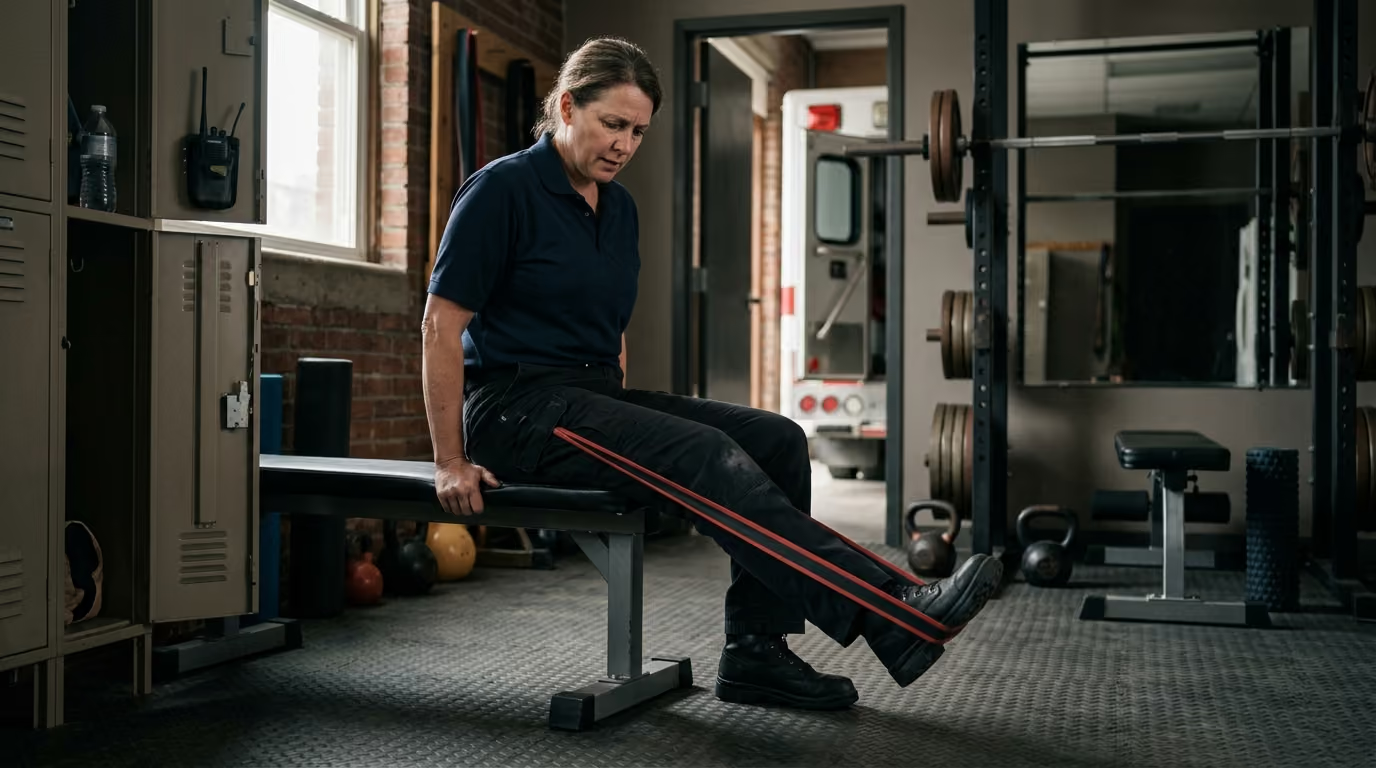
Clinical practice guidelines for midportion Achilles tendinopathy recommend progressive mechanical loading as the first-line intervention. Tendons require high-load, slow-velocity resistance exercise to stimulate collagen remodeling and increase tensile strength. Complete rest weakens the tendon structure, making it more vulnerable to reinjury when explosive tasks are resumed.
Progression along this continuum requires monitoring functional benchmarks. Sports medicine research suggests that daily walking pain should remain at or below a 2 out of 10 before initiating running drills. Furthermore, performing a 20-hop test on one leg without exceeding a 2 out of 10 pain rating provides an objective indicator that the tendon can tolerate dynamic impact.
If a responder feels a sudden pop in the calf, loses the ability to push off the foot, or discovers a palpable gap in the tendon, they must treat the injury as an acute rupture. Ruptures require immediate clinical evaluation rather than progressive exercise loading.
Returning to unrestricted duty requires more than just waiting for pain to subside. The United States Olympic and Paralympic Committee (USOPC) tendinopathy guidelines emphasize that symptom resolution alone does not prove an individual is ready for high-demand activity. A responder may feel comfortable during everyday tasks while remaining underprepared for the chaotic, heavy physical demands of emergency response.
Operational readiness must be assessed across four distinct domains: symptoms, capacity, skill, and resilience. Physical testing should progress from isolated gym exercises to integrated duty simulations conducted while wearing personal protective equipment.
A firefighter recovering from knee irritation should follow a graded progression to verify physical capacity before returning to full structural firefighting duties.
Each phase should be evaluated using the 24-hour pain-monitoring rule. The responder advances to the next phase only when the current tier is completed with clean movement mechanics and no next-morning symptom spike.
An emergency medical technician or paramedic recovering from a lower back flare-up requires a structured return progression centered around patient handling and confined-space management.
Progressing through these duty-specific milestones ensures that the provider's physical capacity matches the unpredictable demands of patient transport. Combining physical progressions with structured recovery and appropriate night duty nutrition supports sustained career longevity.
Station culture often perpetuates myths regarding injury management and strength training. Correcting these misconceptions helps personnel make evidence-informed decisions about their physical health.
The belief that all painful conditions require total rest is outdated. While acute fractures and structural tears need initial immobilization, non-traumatic joint and tendon issues deteriorate with prolonged bed rest. Inactivity leads to muscle atrophy, reduced tendon stiffness, and heightened central nervous system sensitivity.
NICE guidelines for low back pain explicitly emphasize remaining active and engaging in tailored exercise once serious conditions are ruled out. The goal is to modify the provoking movement while maintaining overall physical activity.
Many responders assume that any sensation of pain indicates active tissue destruction. Pain is an alarm system modulated by systemic factors, including sleep deprivation, psychological stress, and physical fatigue.
An acute flare-up often reflects tissue sensitivity and temporary inflammation rather than new structural tearing. Understanding this distinction prevents fear-avoidance behaviors that lead to unnecessary deconditioning.
Responders frequently demand immediate magnetic resonance imaging (MRI) or X-rays when experiencing joint discomfort. While imaging is essential for diagnosing traumatic injuries, fractures, and suspected malignancies, it is poorly correlated with non-traumatic pain.
Degenerative disc changes, meniscus fraying, and rotator cuff fraying are common in asymptomatic, highly active individuals. Relying strictly on imaging findings can cause unnecessary anxiety and lead to unneeded interventions. Training adjustments should be guided by functional movement capacity rather than an isolated scan report.
When a joint aches, personnel often assume the surrounding muscles are tight and require aggressive stretching. However, stretching an irritated tendon or inflamed joint capsule can worsen symptoms by applying compressive and tensile stress at end-range angles. Tendons and joints typically respond far better to graded resistance training, isometric holds, and load management than to prolonged static stretching.
Lifting heavy weights in the gym does not automatically guarantee safe duty performance. A responder who can deadlift heavy weight off a flat barbell platform may still struggle to carry a heavy patient down a narrow, winding staircase while fatigued. Operational capability requires movement control, multi-planar stability, work capacity, and the ability to maintain clean lifting mechanics when exhausted. Training around pain must prioritize functional resilience over raw gym numbers.
When joint or tendon pain appears during a training cycle, responders can execute a simple, five-step algorithm to navigate the problem safely.
Begin by ruling out conditions that require immediate medical attention. Check for acute trauma, visible joint deformity, massive swelling, inability to bear weight, severe neurological symptoms, and systemic signs of illness. If any of these red flags are present, stop training and seek immediate medical evaluation.
If red flags are absent, analyze the training session to identify what triggered the irritation. Record the specific exercise, the working load, the total sets and repetitions, the movement velocity, and the fatigue state. Consider external contributors, such as recent shift length, sleep debt, and demanding calls handled earlier in the week.
Make targeted adjustments to the provoking exercise. Start by reducing the external load by 10% to 30%, shortening the range of motion, or slowing down the movement tempo. If the movement remains uncomfortable, substitute a less provocative variation that trains the same pattern, such as switching from a barbell press to a landmine press. Avoid changing every exercise in the program simultaneously; adjust one or two variables so you can measure what works.
Use the pain-monitoring model to evaluate how the modified movement is tolerated. Ensure discomfort remains at or below a 5 out of 10 during the session and returns to pre-training baseline by the next morning. If symptoms remain stable or improve over seven days, maintain the current dose and progress gradually. If baseline pain increases, reduce the training stress further.
If joint or tendon pain persists for more than two to three weeks despite structured modifications, arrange an evaluation with a qualified sports physical therapist or occupational healthcare provider. Professional assessment is also necessary if pain is accompanied by joint instability, progressive strength loss, or radiating nerve pain that interferes with regular duty tasks. A clinical examination provides clarity, rules out serious structural pathology, and establishes a clear path for rehabilitation.
To see this algorithm in action, consider how it applies to common operational situations.
Applying a structured, evidence-aware system ensures that physical setbacks are managed logically. By treating pain as actionable feedback rather than an excuse for total inactivity, first responders can maintain physical readiness, protect their health, and build lasting career capability.
Maintaining physical capability throughout an emergency service career requires treating pain with practical, evidence-based adjustments that protect operational readiness and long-term health.
Stay connected for research-led guidance on duty fitness, shift-work sleep, injury recovery, stress, nutrition and career longevity. Practical ideas for first responders who want to stay capable on the job, recover well and protect their health for the long run.



Read practical guidance on first responder fitness, recovery, sleep, nutrition and resilience for demanding work and a long, active career.
read the blog