
Feeling strong after weeks of conditioning masks the slower adaptation of tendons, requiring emergency personnel to follow staged progressive loading protocols for lasting joint durability.





A persistent ache at the front of the knee during stair climbs, morning stiffness in the Achilles tendon, or sharp elbow pain when gripping tools often prompts emergency personnel to search for answers. These symptoms rarely stem from sudden muscle weakness or bad luck. They usually reflect a mismatch between the mechanical capacity of your tendons and the sudden spikes in physical load demanded by emergency service. This guide provides a definitive resource on tendon biology, occupational strain, progressive loading protocols, and recovery management for sustained operational readiness.
Tendons require weeks to months of progressive mechanical tension to alter their material stiffness and load capacity. Muscle tissue adapts to training loads much faster than dense connective tissue. When emergency calls demand maximum physical output after quiet station periods, underprepared tendons absorb forces they cannot yet tolerate. Rebuilding tendon resilience requires structured, heavy, and slow mechanical loading combined with strict workload management rather than passive rest.
A tendon is a specialized fibrous connective tissue that connects muscle to bone. Its primary role is transmitting mechanical force across joints to produce movement, stabilize joint positions, and store elastic energy. The internal architecture of a tendon consists predominantly of parallel bundles of type I collagen fibers embedded within a ground substance matrix. Tenocytes, the resident tendon cells, sense mechanical forces placed on the tissue through a process called mechanotransduction. When mechanical tension stretches these cells, they initiate biochemical signaling cascades that synthesize new collagen and remodel the surrounding matrix.
Chronic mechanical loading alters three distinct tendon properties over time. These properties include stiffness, material modulus, and cross-sectional area. Stiffness describes the amount of force required to elongate the tendon by a specific distance. Modulus represents the material stiffness of the tissue independent of its physical dimensions. Cross-sectional area refers to the physical thickness of the tendon structure. Research shows that high-magnitude mechanical loading produces reliable increases in tendon stiffness and tissue modulus, along with modest increases in cross-sectional area.
Tendon adaptation operates on a completely different timeline than muscle tissue. Skeletal muscle possesses an abundant capillary blood supply and high metabolic turnover, allowing it to adapt to training stressors within a few weeks. Dense regular connective tissue receives significantly lower vascular perfusion. Oxygen consumption in tendons is roughly seven times lower than in skeletal muscle. Consequently, the cellular synthesis and cross-linking of collagen fibers require months of consistent, appropriately dosed mechanical tension.
This biological timeline creates a common clinical problem in physical training. A first responder may feel strong, energetic, and muscularly capable after four weeks of aggressive conditioning. However, the tendons attaching those muscles to bone have barely begun their structural remodeling process. If training volume, running speed, or occupational lifting increases rapidly during this window, the tendon experiences microtrauma faster than its tenocytes can repair the matrix. Understanding this discrepancy prevents premature progression and protects against chronic degenerative changes.
Symptom changes must also be distinguished from true structural adaptation. An isometric hold or a brief period of modified activity can reduce pain within minutes by altering local nervous system sensitivity. Medium-term capacity improvements, such as increased muscular endurance and improved movement tolerance, typically emerge over six to twelve weeks. Long-term tissue remodeling, including measurable increases in tendon thickness and material stiffness, requires several months of sustained loading. Feeling pain-free during a single shift does not mean the tendon has finished remodeling.
First responders operate under a unique physical paradigm characterized by low baseline activity punctuated by sudden, maximal occupational peaks. Firefighters, police officers, and paramedics spend hours sitting in vehicles, completing paperwork, or performing low-intensity station tasks. Without warning, an emergency dispatch requires immediate high-output physical exertion. This shift from resting state to maximal physical exertion places severe, unprimed tensile and compressive forces on tendon structures.
Musculoskeletal disorders represent a massive occupational burden across emergency services. Research indicates that the pooled prevalence of musculoskeletal disorders among firefighters reaches approximately 41 percent. Regional pain reports show high vulnerability across multiple joints. Shoulder pain exhibits point prevalence rates near 23 percent, knee pain reaches 27 percent, and general sprains and strains account for a large portion of all documented firefighter injuries.
Law enforcement personnel experience similar patterns of soft-tissue strain, particularly in the upper extremities. Physical encounters with uncooperative subjects, defensive tactics training, and tactical shooting drills place intense, erratic loads on the wrist, elbow, and shoulder joints. Systematic reviews of police officers demonstrate 12-month symptom frequencies reaching up to 27 percent for the wrist and hand, 10 percent for the elbow, 32 percent for the shoulder, and 28 percent for the ankle and foot. Soft-tissue sprains, strains, and tendinopathies occur repeatedly across patrol and tactical units alike.
External equipment substantially magnifies occupational joint and tendon loading. Firefighters carry between 45 and 75 pounds of personal protective equipment, breathing apparatus, and hand tools. Tactical law enforcement officers wear body armor, duty belts, and tactical gear that alter their center of mass and gait mechanics. Paramedics lift bariatric patients using manual stretchers in narrow stairwells and confined spaces. These loads alter normal movement paths, increase ground reaction forces during running or stair climbing, and force tendons to manage heavy tensile loads while simultaneously enduring bone-on-tendon compression.
Occupational movement in emergency services is inherently unpredictable. Unlike controlled gym environments where a barbell moves along a predictable vertical path, emergency tasks involve dynamic, multi-planar resistance. A charged hose line kicks back with shifting water pressure. A patient may shift weight or drop suddenly during a stair-chair transfer. A combative individual actively resists physical control. These uncontrolled perturbations generate sudden rates of force application that challenge tendon elasticity and demand robust mechanical tolerance.
Physical loading on a tendon is not defined solely by the external weight listed on a barbell. Tendon stress is multi-dimensional and involves ten distinct, interacting variables:
Workload spikes represent the single greatest modifiable risk factor for tendon injury. A workload spike occurs when acute physical demands significantly outpace the chronic workload the individual has prepared for over the preceding four to six weeks. In athletic populations, large weekly increases in external workload consistently precede muscle and tendon breakdowns. First responders encounter severe workload spikes during academy recruit training, after returning from extended medical or vacation leave, during sudden staffing shortages, or after consecutive mandatory overtime shifts.
Energy-storage loading carries the highest risk for tendon irritation. Activities such as sprinting, jumping down from apparatus cabs, rapid stair descent, and explosive directional changes require tendons to act like mechanical springs. The Achilles and patellar tendons quickly store kinetic energy and release it within fractions of a second. While slow, heavy resistance training builds the foundational force capacity of the muscle-tendon unit, it does not fully prepare the tendon for rapid elastic deformation. If high-velocity energy-storage tasks are introduced without progressive preparation, reactive tendon symptoms frequently follow.
Compressive loading represents another critical mechanism of tendon irritation. Tendons experience tensile stress along their longitudinal fibers, but they also experience compressive stress when wrapped tightly around a bony prominence at end-range joint angles. For example, deep squatting or kneeling compresses the patellar and quadriceps tendon insertions against the femoral condyles. Full wrist extension under load compresses the wrist flexor tendons within the carpal canal. Sustained shoulder elevation compresses the supraspinatus tendon beneath the acromion arch. Combining heavy tensile resistance with extreme compressive angles creates high shear stress that provokes localized tendon pathology.
To balance systemic physical readiness with duty recovery, responders can review protocols for programming high output capacity workouts alongside low fatigue maintenance sessions across varying shift schedules.
Progressive therapeutic loading serves as the primary, evidence-supported intervention for improving tendon capacity and resolving tendinopathy. Complete rest removes the vital mechanical stimulus required for collagen synthesis, leading to tissue stress shielding and further capacity reduction. Conversely, aggressive, unmonitored training aggravates inflamed or degenerated tendon regions. A structured, four-stage progressive loading framework bridges this gap by systematically expanding tissue tolerance.
When a tendon is irritable and reactive, dynamic movement often causes sharp pain and reactive swelling. Heavy isometric contractions provide a valuable entry point. Static muscular contractions performed at mid-range joint angles can reduce localized tendon pain through exercise-induced hypoalgesia while maintaining motor unit recruitment. Clinical consensus panels support isometric loading as an effective method for immediate symptom management when dynamic movements are poorly tolerated.
A standard isometric loading protocol utilizes four to five sets of 30- to 45-second holds at roughly 70 percent of maximal voluntary effort. The joint angle should be chosen to minimize compressive impingement. For the patellar tendon, Spanish squats or single-leg leg extensions held at 60 degrees of knee flexion work well. For the Achilles tendon, seated or standing mid-range calf holds provide stable tension. For lateral elbow tendinopathy, isometric wrist extension held in neutral alignment reduces local pain.
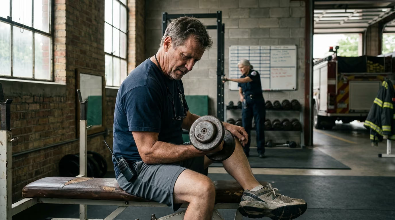
Once resting irritability subsides, the primary training objective shifts to building mechanical tissue stiffness, muscle hypertrophy, and load tolerance. Heavy Slow Resistance (HSR) utilizes high external loads moved through controlled ranges of motion with extended time under tension. Research confirms that heavy isotonic loading at 70 to 80 percent of one-repetition maximum produces significant increases in tendon stiffness and material modulus over 12-week training cycles.
A typical HSR protocol involves three to four sets of six to eight repetitions, with each repetition lasting six to eight seconds total (three to four seconds concentric, three to four seconds eccentric). This slow cadence eliminates momentum, minimizes sudden rate-of-force spikes, and ensures uniform mechanical strain across the tendon fibers. Exercises should be performed three times per week on non-consecutive days to allow sufficient collagen turnover and matrix recovery between bouts.
A tendon can be strong under slow speeds yet remain completely unprepared for explosive occupational tasks. Once heavy slow resistance is tolerated without reactive flare-ups, dynamic energy-storage and release exercises must be introduced. This phase systematically restores the stretch-shortening cycle capability of the tendon unit.
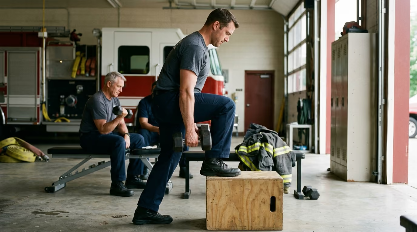
Progression begins with low-amplitude, controlled elastic tasks. For lower extremity tendons, this includes two-legged ankle hops, skipping rope, and low-height box step-downs. As tolerance develops, training advances to single-leg hopping, broad jumps, rapid change-of-direction shuttle runs, and loaded stair descents. Upper extremity progressions include medicine ball chest passes, explosive cable pulls, and rapid catch-and-release drills. Volume must start low, and sessions should be separated by at least 48 to 72 hours.
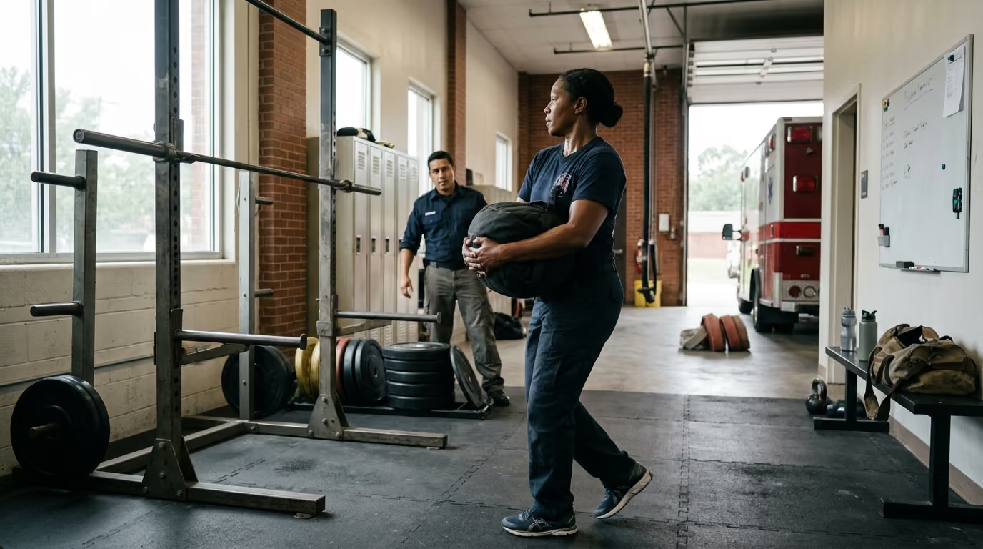
The final phase bridges athletic capacity to operational duty requirements. Controlled gym movements cannot fully simulate dragging a 200-pound victim, advancing a charged hoseline through a doorway, or restraining a combative subject on uneven ground. Stage 4 introduces multi-planar instability, equipment weight, and mild physical fatigue into functional task rehearsals.
Training tasks in this stage include dummy drags wearing weighted vests, structural tool strikes against resistance tires, sled pushes mimicking vehicle extrication maneuvers, and partner grappling drills. These tasks must be dosed deliberately. Responders should never introduce multiple new high-demand operational drills during the same week they face heavy shift-work overtime.
For a broader perspective on tissue management and physical longevity across a full career, visit our collection on injury prevention and physical recovery.
Different anatomical regions experience distinct mechanical challenges based on joint architecture and occupational duties. Structuring targeted exercises for each major tendon group builds specific local capacity.
The extensor mechanism of the knee absorbs massive kinetic energy during crawling, crouching in confined spaces, descending stairs, and landing from apparatus steps. The patellar tendon connects the inferior pole of the patella to the tibial tuberosity, enduring extreme tensile loads during knee flexion under load.
Programming priorities for the patellar tendon include:
The Achilles tendon is the thickest and strongest tendon in the human body, transmitting forces up to six to eight times body weight during running and jumping. Heavy structural bunker boots restrict normal ankle dorsiflexion, shifting mechanical stress directly into the mid-portion and insertion of the Achilles tendon.
Programming priorities for the Achilles tendon include:
Shoulder tendons, particularly the supraspinatus and the long head of the biceps, operate in tight anatomical spaces. Firefighters working with pike poles on ceilings, paramedics transferring heavy stretchers, and officers controlling subjects subject these tendons to high tensile strain and subacromial compression.
Programming priorities for the shoulder complex include:
The common extensor tendon at the lateral epicondyle and the common flexor tendon at the medial epicondyle manage all forceful grip and wrist stabilization tasks. Operating hydraulic extrication tools, carrying heavy medical jump bags, and applying joint locks place intense demands on these forearm muscle-tendon units.
Programming priorities for the elbow and wrist include:
For structured guidance on minimal training doses to keep these joints operational during intense shift rotations, refer to our framework on balancing structured physical training and short readiness sessions.
Managing tendon load requires an ongoing feedback loop rather than rigid adherence to a static calendar schedule. Tendons often fail to register pain during the actual training session because local warming increases tissue compliance. Pain and stiffness frequently manifest 12 to 24 hours later, once local biochemical inflammatory mediators accumulate.
The 24-hour response serves as the primary gauge for evaluating whether a physical load was appropriate. First responders should track pain levels during activity and assess local stiffness the following morning. Research on lower limb tendinopathy indicates that mild to moderate pain (up to 4 or 5 out of 10 on a numeric pain rating scale) is acceptable during and immediately after exercise. However, two non-negotiable criteria must be met:
A practical traffic-light monitoring system helps operational personnel make objective daily adjustments to their training and duty preparation:
Tendon capacity is heavily modified by systemic recovery factors. Sleep deprivation, long shift rotations, acute mental stress, and poor nutritional intake impair systemic collagen synthesis and lower the mechanical threshold at which a tendon becomes symptomatic. If an emergency responder works a brutal 24-hour shift with zero sleep, their tendon load tolerance the following day is significantly depressed. Training loads must be autoregulated to match the individual's systemic readiness rather than forced through accumulated fatigue.
To understand how consecutive shifts and interrupted sleep impair musculoskeletal recovery pathways, examine the analysis on how shift rotations drive fatigue and sleep debt.
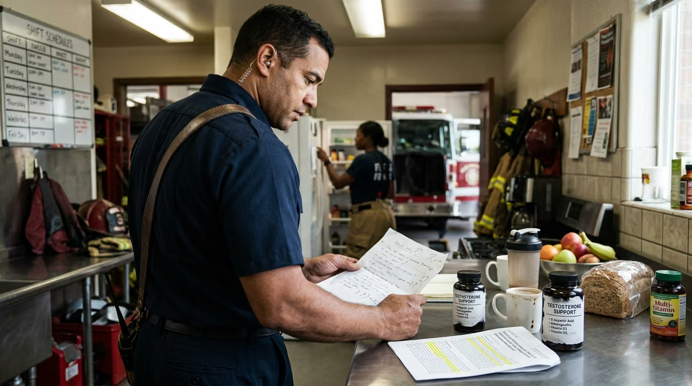
Tendon rehabilitation cannot be solved by mechanical exercise alone if the biological environment within the body is compromised. Tendon tissue is metabolically active and sensitive to systemic health, hormonal balance, and nutritional status. Recent clinical research emphasizes screening for metabolic dysregulation in persistent, treatment-resistant tendinopathies.
Metabolic conditions such as insulin resistance, dyslipidemia, systemic low-grade inflammation, and elevated visceral adiposity compromise tendon microcirculation. Advanced glycation end-products accumulate within the collagen matrix in individuals with elevated blood glucose levels. This accumulation causes abnormal, brittle cross-linking between collagen fibers, reducing overall tendon elasticity and increasing susceptibility to microtrauma under normal operational loads.
Dietary protein intake provides the basic amino acid building blocks required for collagen synthesis and extracellular matrix repair. Active first responders should consume 1.6 to 2.2 grams of protein per kilogram of body weight daily. This protein intake should be distributed across three to five meals containing high-quality essential amino acids, including leucine, proline, and glycine. Hydration also plays a key role, as the proteoglycan ground substance within tendons relies on bound water molecules to maintain compressive cushioning and smooth inter-fascicular sliding.
While dietary supplements such as hydrolyzed collagen peptides and vitamin C receive substantial marketing attention, the scientific evidence supporting their independent efficacy remains preliminary and mixed. Ingesting 15 to 20 grams of collagen or gelatin along with 50 milligrams of vitamin C roughly 45 to 60 minutes before loading may support collagen synthesis markers in laboratory settings. However, these supplements serve only as minor adjuncts. They cannot replace properly programmed mechanical loading, adequate sleep, and total caloric sufficiency.
Understanding how sleep deficits from shift work accelerate structural breakdown can be explored further in the review of CDC baselines for shift-work fatigue and recovery.
While progressive loading effectively addresses uncomplicated tendinopathy, first responders must recognize when local symptoms indicate a more severe pathology. Pain around a joint does not automatically equal simple tendon irritation. Bone stress injuries, joint labral tears, ligamentous ruptures, nerve entrapments, and acute partial or complete tendon tears require immediate, specialized medical intervention.
Clear clinical red flags require immediate cessation of training and prompt evaluation by a licensed healthcare professional:
Self-treating an undiagnosed condition through aggressive loading can turn a minor partial tear into a catastrophic complete rupture requiring surgical repair. Returning to active duty while limping, guarding a joint, or lacking grip strength compromises personal safety and places fellow crew members at severe operational risk during critical emergency events.
For specialized clinical insights into severe occupational physical trauma, read our resource on specialized rehabilitation and testing protocols for emergency personnel. Readers can also explore our broader directory of first responder injury and recovery guides for detailed joint protocols.
Building occupational tendon resilience requires coordinated action across both individual first responders and administrative departments. Organizations must move past reactive injury management and adopt proactive workload monitoring systems that preserve frontline operational capability.
Departments often witness large injury waves when personnel encounter sudden operational surges after prolonged quiet seasons or administrative assignments. Maintaining an adequate physical baseline (the capacity "floor") prevents severe workload spikes when high-demand incidents occur. Station routines should incorporate year-round, low-volume maintenance sessions focusing on calf stiffness, grip endurance, rotator cuff control, and multi-planar core stability.
The maximum physical demand of the job (the operational "ceiling") must be approached systematically. Academy training programs and department fitness coordinators must avoid exposing unconditioned recruits or returning personnel to full-gear, high-speed, multi-mile evolutions on day one. Running, stair climbs, heavy carries, and live-fire drills should be layered across multiple weeks. This gives dense connective tissues adequate time to remodel between progressive exposures.
When designing station conditioning or recruit physical training, never increase multiple stress variables simultaneously. If you increase running speed or stair climbing cadence, maintain or reduce carried equipment weight. If you introduce heavy bunker gear and tools, reduce total repetition volume and step distance. Changing only one variable at a time makes tissue tolerance predictable and prevents acute loading spikes.
Returning an injured responder to duty based purely on an arbitrary calendar timeline (such as "six weeks of light duty") frequently results in immediate reinjury. Tendon remodeling does not adhere to standard administrative calendars. Clearance to full duty should require passing objective functional tests:
To apply these principles directly to your routine this week, work through the following checklist:
Building tendon resilience is a continuous, methodical process that requires patience, controlled mechanical load, and disciplined recovery tracking. Treating your connective tissues with the same structured focus as cardiovascular conditioning ensures a long, capable career free from debilitating joint breakdowns.
Stay connected for research-led guidance on duty fitness, shift-work sleep, injury recovery, stress, nutrition and career longevity. Practical ideas for first responders who want to stay capable on the job, recover well and protect their health for the long run.



Read practical guidance on first responder fitness, recovery, sleep, nutrition and resilience for demanding work and a long, active career.
read the blog